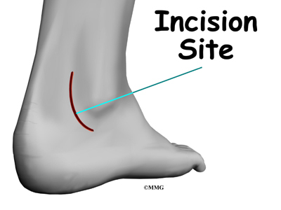
Torn Retinaculum : Diagram Showing The Position Of The Skin Incision For Approach To The Download Scientific Diagram / Tearing of retinacula is more commonly seen at the ankle.
byAdmin•
0
Torn Retinaculum : Diagram Showing The Position Of The Skin Incision For Approach To The Download Scientific Diagram / Tearing of retinacula is more commonly seen at the ankle.. When the knee moves slightly out of place or becomes tilted in the joint, it can cause tension and pain in the lateral retinaculum. The skin is closed with stitches. Tear of the superior peroneal retinaculum at its attachment to the distal fibula Tearing of retinacula is more commonly seen at the ankle. The examiner can usually feel the gap in the tendon, just below the kneecap.
The tear is repaired with suture above. Tendonitis is an inflammation of one or both tendons. Stretching this ligament keeps the patella in place and the ligament healthy.stretching a lateral retinaculum of the knee. Tendon tear is repaired with suture and tendon is returned to tubular shape. Previously torn extensor retinaculum of ankle which is now markedly thickened and irregular (blue arrows).
Patient Education Concord Orthopaedics from www.eorthopod.com The flexor retinaculum of the foot can be strained or injured due to a variety of reasons. It thickens as it inserts onto the. The skin is closed with stitches. Sportsmen involved in running and sprinting can strain or inflame this structure. The medial and lateral patellar retinaculum are on their respective sides of the patella and are continuous with the vastus fascia to the tibia and the patella. Making the diagnosis of a torn patellar tendon is usually obvious on clinical examination. Between 2001 and 2011 three patients with distal peroneal tendon dislocation were operated. The patella is a sesamoid bone.
The torn edge of the retinaculum is then pulled into the trough and sutured in place.
Occasionally, the covering that holds the peroneus tendons behind the lateral malleolus (the retinaculum) can be loose or torn and the tendons can snap back and forth out of their normal grooves, this snapping sensation is felt by the patient and can causes further stress/friction on the tendons. Tearing of retinacula is more commonly seen at the ankle. A popping or tearing sensation at the time of injury. Sportsmen involved in running and sprinting can strain or inflame this structure. There was no tearing in any other part of the capsule of the hip joint in any patient. The surgeon uses a burr to create a trough along the fibula bone next to the original attachment of the retinaculum. Plantaris tendon (if you have one) overlay may do the. People who tear the tendon will be unable to extend their knee against gravity, and unable to perform a straight leg raise test. The first step to regaining full recovery of your range of motion is to quickly identify an injury to the patellar tendon. Tear of the superior peroneal retinaculum at its attachment to the distal fibula Stretching this ligament keeps the patella in place and the ligament healthy.stretching a lateral retinaculum of the knee. Tendonitis is an inflammation of one or both tendons. In addition moving around with a torn meniscus could pull fragments of the cartilage into the joint causing larger knee issues which could.
Between 2001 and 2011 three patients with distal peroneal tendon dislocation were operated. Pain and swelling of the knee. The symptoms of a partial or complete patellar tendon tear may include: Plantaris tendon (if you have one) overlay may do the. The skin is closed with stitches.
Ankle Stability Retinaculum Connection from static.wixstatic.com The retina plays a vital role in vision. When the knee moves slightly out of place or becomes tilted in the joint, it can cause tension and pain in the lateral retinaculum. The patella is a sesamoid bone. The lateral retinaculum is a ligament that helps hold your patella, or kneecap, in place. In this article, we present the assessment, diagnostic algorithm and a new therapeutic option for the distal dislocation of the long peroneal tendon due to isolated inferior peroneal retinaculum (ipr) tear. People who tear the tendon will be unable to extend their knee against gravity, and unable to perform a straight leg raise test. The posterior retinaculum was found torn in two garden type iv fractures. Most of the fibers of the medial patellar retinaculum originate in the medial femoral region from the vastus medialis muscle, just superior to the patella.
In addition moving around with a torn meniscus could pull fragments of the cartilage into the joint causing larger knee issues which could.
Learn more about the types, causes, risk factors, symptoms, diagnosis, treatment. Tear of the superior peroneal retinaculum at its attachment to the distal fibula Tendon tear is repaired with suture and tendon is returned to tubular shape. Sportsmen involved in running and sprinting can strain or inflame this structure. The torn edge of the retinaculum is then pulled into the trough and sutured in place. They are minor patellar stabilizers and, if intact, can provide knee extension and straight leg raising despite a patellar or quadriceps tendon rupture. If the groove at the point of the heel bone (tuber calcanei) is found to be absent or abnormally shallow, it will be deepened to further increase stability. The posterior retinaculum was found torn in two garden type iv fractures. For a torn peroneus brevis tendon, the proximal tenodesis transfers or attaches the brevis to the longus sufficiently proximal to the superior peroneal retinaculum such that the combined tendons at the tenodesis will not pass through the narrow zone of the retinaculum even in maximum inversion. Moderate to severe pain in the ankle and foot with ambulating or running or in fact any movement of the foot. The posterior retinaculum was found intact in all of the garden type iii fractures and in 39 garden type iv fractures. Occasionally, the covering that holds the peroneus tendons behind the lateral malleolus (the retinaculum) can be loose or torn and the tendons can snap back and forth out of their normal grooves, this snapping sensation is felt by the patient and can causes further stress/friction on the tendons. In this article, we present the assessment, diagnostic algorithm and a new therapeutic option for the distal dislocation of the long peroneal tendon due to isolated inferior peroneal retinaculum (ipr) tear.
Left untreated, a meniscus tear can limit your daily life and ability to participate in exercise and sports. A retinal tear can lead to fluid and blood collecting in the eye, which can cause the development of several new floaters and loss of vision if the tear leads to a retinal detachment. Stretching this ligament keeps the patella in place and the ligament healthy.stretching a lateral retinaculum of the knee. Sportsmen involved in running and sprinting can strain or inflame this structure. Tendon tear is repaired with suture and tendon is returned to tubular shape.
Icd 10 Code For Tear Of The Medial Patellar Retinaculum from s-media-cache-ak0.pinimg.com Likely to be caused by a fatigue tear in the medial capsular insertion into the patella (dutton). Sportsmen involved in running and sprinting can strain or inflame this structure. Stretching this ligament keeps the patella in place and the ligament healthy.stretching a lateral retinaculum of the knee. Previously torn extensor retinaculum of ankle which is now markedly thickened and irregular (blue arrows). They are minor patellar stabilizers and, if intact, can provide knee extension and straight leg raising despite a patellar or quadriceps tendon rupture. Most of the fibers of the medial patellar retinaculum originate in the medial femoral region from the vastus medialis muscle, just superior to the patella. When the knee moves slightly out of place or becomes tilted in the joint, it can cause tension and pain in the lateral retinaculum. Tearing of retinacula is more commonly seen at the ankle.
The medial and lateral patellar retinaculum are on their respective sides of the patella and are continuous with the vastus fascia to the tibia and the patella.
Can this be cured without surgery? In addition moving around with a torn meniscus could pull fragments of the cartilage into the joint causing larger knee issues which could. The torn edge of the retinaculum is then pulled into the trough and sutured in place. Previously torn extensor retinaculum of ankle which is now markedly thickened and irregular (blue arrows). Ankle ext retinaculum torn after fasciotomy & injury in pt. The tendon sheaths and retinaculum (structure that binds tendons down) are repaired. Occasionally, the covering that holds the peroneus tendons behind the lateral malleolus (the retinaculum) can be loose or torn and the tendons can snap back and forth out of their normal grooves, this snapping sensation is felt by the patient and can causes further stress/friction on the tendons. The flexor retinaculum of the foot can be strained or injured due to a variety of reasons. The symptoms of a partial or complete patellar tendon tear may include: The skin is closed with stitches. The first step to regaining full recovery of your range of motion is to quickly identify an injury to the patellar tendon. In this article, we present the assessment, diagnostic algorithm and a new therapeutic option for the distal dislocation of the long peroneal tendon due to isolated inferior peroneal retinaculum (ipr) tear. The lateral retinaculum is a ligament that helps hold your patella, or kneecap, in place.